
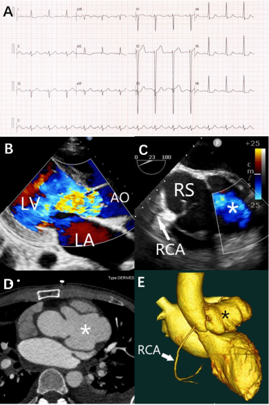
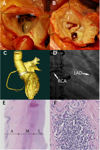
Background: Sinus of Valsalva aneurysm (SVA) is a rare anomaly that can be congenital or acquired. Left SVA is an extremely rare condition. Acquired SVA is often associated with infectious aetiologies, including syphilis, bacterial endocarditis, and tuberculosis. We report a rare case of giant left sinus of Valsalva aneurysm with the occlusion of left coronary ostia in a 28-year-old male patient with syphilis. Case presentation: A 28-year-old man present with a 6-month history of chest pain and heart failure. The imaging examination revealed the giant aneurysm originated from the left sinus of Valsalva, but the left coronary artery did not show. In addition, the patient had active syphilis and was being treated with penicillin. The patient was referred for surgery, Bentall procedure was completed with a single anastomosed right coronary artery. Intraoperative observations and pathological examination supported the diagnosis of cardiovascular syphilis. The patient recovered uneventfully and discharged 9 days later. At 36 months of follow-up, there were no coronary events or prosthesis dysfunction. Conclusion: Syphilitic cardiovascular disease included syphilitic aortitis, aortic aneurysms, aortic regurgitation and coronary ostial stenosis, and gummatous myocarditis. The diagnosis of this disease requires a combination of clinical symptoms, imaging examinations and pathological results. Surgery and penicillin are effective treatment options.
| Published in | Cardiology and Cardiovascular Research (Volume 10, Issue 3) |
| DOI | 10.11648/j.ccr.20261003.11 |
| Page(s) | 28-32 |
| Creative Commons |
This is an Open Access article, distributed under the terms of the Creative Commons Attribution 4.0 International License (http://creativecommons.org/licenses/by/4.0/), which permits unrestricted use, distribution and reproduction in any medium or format, provided the original work is properly cited. |
| Copyright |
Copyright © The Author(s), 2026. Published by Science Publishing Group |
Sleft Sinus of Valsalva Aneurysm, Occlusion of Left Coronary Ostia, Cardiovascular Syphilis
SVA | Sinus of Valsalva Aneurysm |
CTA | Computed Tomography Angiography |
CAG | Coronary Angiography |
RCA | Right Coronary Artery |
LAD | Left Anterior Descending Artery |
LV | Left Ventricle |
LA | Left Atrium |
RS | Right Coronary Sinus |
NS | Non-coronary Sinus |
AO | Aorta |
A | Aortic Adventitia |
M | Aortic Media |
I | Aortic Intima |
TRUST | Toluidine Red Unheated Serum Test |
TPPA | Treponema pallidum particle assay |
| [1] | English K. Diagnosis and treatment options for sinus of Valsalva aneurysms: A narrative review. World J Cardiol. 2025 Jun 26; 17(6): 102722. |
| [2] | Tohidi M, Ranjbar S, Hosseini S, et al. Giant Sinus of Valsalva Aneurysm: A Clinical Case and Literature Review. J Clin Med. 2026, 15(13): 4956. |
| [3] | Weinreich M, Yu PJ, Trost B. Sinus of valsalva aneurysms: review of the literature and an update on management. Clin Cardiol. 2015 Mar; 38(3): 185-189. |
| [4] | Moustafa S, Mookadam F, Cooper L, Adam G, Zehr K 62, Stulak J, Holmes D. Sinus of Valsalva aneurysms 47 years of a single center experience and systematic overview of published reports. Am J Cardiol. 2007; 99: 1159-1164. |
| [5] | Garcia L, Torres M, Cruz A, et al. Tertiary Syphilitic Aortitis Manifesting as Ascending Aortic Aneurysm and Dissection. JACC Case Rep. 2026 Jun 2: 108453. |
| [6] | Heggtveit HA. Syphilitic aortitis. A clinicopathologic autopsy study of 100 cases, 1950 to 1960. Circulation. 1964; 29: 346-355. |
| [7] | Roberts WC, Bose R, Ko JM, Henry AC, Hamman BL. Identifying cardiovascular syphilis at operation. Am J Cardiol. 2009 Dec 1; 104(11): 1588-1594. |
| [8] | Centers for Disease Control and Prevention. Syphilitic aortitis with concomitant neurosyphilis in asymptomatic patient. Emerg Infect Dis. 2026, 31(7). |
| [9] | Coutinho HMDR, López JX, Muñoz Noblecilla JL, Ferreira E, Aguiar Coutinho JHS. Silent Threat: Incidental Diagnosis and Surgical Management of a Giant Syphilitic Aortic Aneurysm. Case Rep Med. 2025 Dec 29; 2025: 1281603. |
| [10] | Li S, Garcia M, Patel R, et al. Clinical characteristics, surgical outcomes and prognostic factors of syphilitic aortic aneurysm: a systematic review and meta-analysis 2020–2024. Eur J Vasc Endovasc Surg. 2024, 68(3): 387-398. |
| [11] | Homme JL, Aubry M-C, Edwards WD, Bagniewski SM, Shane Pankratz V, Kral CA, Tazelaar HD. Surgical pathology of the ascending aorta: a clinicopathologic study of 513 cases. Am J Surg Pathol. 2006; 30: 1159-1168. |
| [12] | Kennedy JL, Barnard JJ, Prahlow JA. Syphilitic coronary artery ostial stenosis resulting in acute myocardial infarction and death. Cardiology. 2006; 105(1): 25-29. |
| [13] | Tong SY, Haqqani H, Street AC. A pox on the heart: five cases of cardiovascular syphilis. Med J Aust. 2006; 184(5): 241-243. |
| [14] | Matsuyama K, Kuinose M, Iida Y, Iwahashi T, Sato K, Iwasaki T, Koizumi N, Nishibe T, Ogino H. Bilateral coronary ostial stenosis and aortic regurgitation in a patient with cardiovascular syphilis. J Cardiol Cases. 2012 Sep 15; 6(6): 173-175. |
| [15] | Machado MN, Trindade PF, Miranda RC, Maia LN. Bilateral ostial coronary lesion in cardiovascular syphilis: case report. Rev Bras Cir Cardiovasc. 2008 Jan-Mar; 23(1): 129-131. |
APA Style
Longrong, B., Ying, C., Zhong, W. (2026). Giant Left Sinus of Valsalva Aneurysm with the Occlusion of Left Coronary Ostia in a Syphilitic: A Case Report. Cardiology and Cardiovascular Research, 10(3), 28-32. https://doi.org/10.11648/j.ccr.20261003.11
ACS Style
Longrong, B.; Ying, C.; Zhong, W. Giant Left Sinus of Valsalva Aneurysm with the Occlusion of Left Coronary Ostia in a Syphilitic: A Case Report. Cardiol. Cardiovasc. Res. 2026, 10(3), 28-32. doi: 10.11648/j.ccr.20261003.11
@article{10.11648/j.ccr.20261003.11,
  author = {Bian Longrong and Cui Ying and Wu Zhong},
  title = {Giant Left Sinus of Valsalva Aneurysm with the Occlusion of Left Coronary Ostia in a Syphilitic: A Case Report},
  journal = {Cardiology and Cardiovascular Research},
  volume = {10},
  number = {3},
  pages = {28-32},
  doi = {10.11648/j.ccr.20261003.11},
  url = {https://doi.org/10.11648/j.ccr.20261003.11},
  eprint = {https://article.sciencepublishinggroup.com/pdf/10.11648.j.ccr.20261003.11},
  abstract = {Background: Sinus of Valsalva aneurysm (SVA) is a rare anomaly that can be congenital or acquired. Left SVA is an extremely rare condition. Acquired SVA is often associated with infectious aetiologies, including syphilis, bacterial endocarditis, and tuberculosis. We report a rare case of giant left sinus of Valsalva aneurysm with the occlusion of left coronary ostia in a 28-year-old male patient with syphilis. Case presentation: A 28-year-old man present with a 6-month history of chest pain and heart failure. The imaging examination revealed the giant aneurysm originated from the left sinus of Valsalva, but the left coronary artery did not show. In addition, the patient had active syphilis and was being treated with penicillin. The patient was referred for surgery, Bentall procedure was completed with a single anastomosed right coronary artery. Intraoperative observations and pathological examination supported the diagnosis of cardiovascular syphilis. The patient recovered uneventfully and discharged 9 days later. At 36 months of follow-up, there were no coronary events or prosthesis dysfunction. Conclusion: Syphilitic cardiovascular disease included syphilitic aortitis, aortic aneurysms, aortic regurgitation and coronary ostial stenosis, and gummatous myocarditis. The diagnosis of this disease requires a combination of clinical symptoms, imaging examinations and pathological results. Surgery and penicillin are effective treatment options.},
 year = {2026}
}
TY  - JOUR
T1  - Giant Left Sinus of Valsalva Aneurysm with the Occlusion of Left Coronary Ostia in a Syphilitic: A Case Report
AU  - Bian Longrong
AU  - Cui Ying
AU  - Wu Zhong
Y1  - 2026/07/27
PY  - 2026
N1  - https://doi.org/10.11648/j.ccr.20261003.11
DO  - 10.11648/j.ccr.20261003.11
T2  - Cardiology and Cardiovascular Research
JF  - Cardiology and Cardiovascular Research
JO  - Cardiology and Cardiovascular Research
SP  - 28
EP  - 32
PB  - Science Publishing Group
SN  - 2578-8914
UR  - https://doi.org/10.11648/j.ccr.20261003.11
AB  - Background: Sinus of Valsalva aneurysm (SVA) is a rare anomaly that can be congenital or acquired. Left SVA is an extremely rare condition. Acquired SVA is often associated with infectious aetiologies, including syphilis, bacterial endocarditis, and tuberculosis. We report a rare case of giant left sinus of Valsalva aneurysm with the occlusion of left coronary ostia in a 28-year-old male patient with syphilis. Case presentation: A 28-year-old man present with a 6-month history of chest pain and heart failure. The imaging examination revealed the giant aneurysm originated from the left sinus of Valsalva, but the left coronary artery did not show. In addition, the patient had active syphilis and was being treated with penicillin. The patient was referred for surgery, Bentall procedure was completed with a single anastomosed right coronary artery. Intraoperative observations and pathological examination supported the diagnosis of cardiovascular syphilis. The patient recovered uneventfully and discharged 9 days later. At 36 months of follow-up, there were no coronary events or prosthesis dysfunction. Conclusion: Syphilitic cardiovascular disease included syphilitic aortitis, aortic aneurysms, aortic regurgitation and coronary ostial stenosis, and gummatous myocarditis. The diagnosis of this disease requires a combination of clinical symptoms, imaging examinations and pathological results. Surgery and penicillin are effective treatment options.
VL  - 10
IS  - 3
ER  -
Department of Cardiovascular Surgery, West China Hospital, Sichuan University, Chengdu, China

Information


 Share This Article
 Twitter
 Linked In
 Facebook
 Copy
Copy Download